
- 3D printing,
- 3D printed Titanium,
- Bone implants,
- hydroxyapatite coatings,
- electrodeposition,
- osteoblast growth
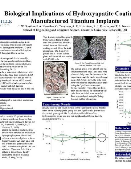
This study sought to determine the growth of and viability of osteoblast cells on hydroxyapatite coatings of 3D-printed titanium implants. The experiment used twenty 3D-printed titanium disks each of which had a determined surface roughness. These disks were printed by Tangible Solutions, LLC (Fairborn, OH) and then sonicated. Ten of the disks were coated with hydroxyapatite through the process of electrodeposition using an open cell and a three lead potentiostat.
Using the hydroxyapatite coated titanium disks and uncoated disks, two four-day growth trials were performed. Two trials used five control disks (uncoated titanium disks) and five coated disks each, making an n of 10 for the total experiment. The disks were each placed into the wells of a culture plate and each disk was seeded with 15,000 human osteoblast cells. After four days in the incubator, the cells were removed using trypsin and the counted using the CytoSmart Automated Hemocytometer. The cell count from each disk as well as the viability of the cells from each disk were recorded. Means comparison was performed using Tukey-Kramer method of analysis.
Results from the cell count portion of the experiment showed that the mean of the hydroxyapatite group was not significantly greater than the control group (p=0.83). In addition, cell viability of the hydroxyapatite group was also not significantly different than the control group (p=0.31). This data was unexpected but may be due to a change in the surface roughness between the two groups caused by the hydroxyapatite coating decreasing the surface roughness. The surface roughness selected for the experiment was chosen due to it being the most ideal for osteoblast growth, but any less rough surfaces were shown to be less ideal for osteoblast cell growth. Future Experiments will remove the variable of surface roughness.
Available at: http://works.bepress.com/timothy_norman/220/