
Article
A unique MRI presentation of fungal infection in the brain
Journal of the College of Physicians & Surgeons of Pakistan
Publication Date
11-1-2014
Document Type
Article
Disciplines
Abstract
ABSTRACT
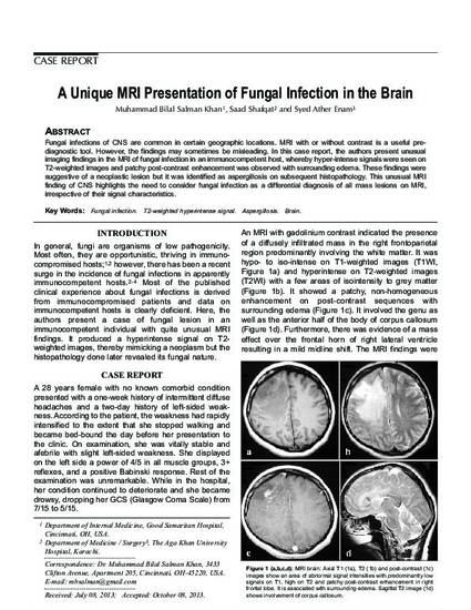
Fungal infections of CNS are common in certain geographic locations. MRI with or without contrast is a useful prediagnostic
tool. However, the findings may sometimes be misleading. In this case report, the authors present unusual
imaging findings in the MRI of fungal infection in an immunocompetent host, whereby hyper-intense signals were seen on
T2-weighted images and patchy post-contrast enhancement was observed with surrounding edema. These findings were
suggestive of a neoplastic lesion but it was identified as aspergillosis on subsequent histopathology. This unusual MRI
finding of CNS highlights the need to consider fungal infection as a differential diagnosis of all mass lesions on MRI,
irrespective of their signal characteristics.
Citation Information
Muhammad B S Khan, Saad Shafqat and Syed A Enam. "A unique MRI presentation of fungal infection in the brain" Journal of the College of Physicians & Surgeons of Pakistan Vol. 24 Iss. SS3 (2014) p. S211 - S213 Available at: http://works.bepress.com/saad_shafqat/5/