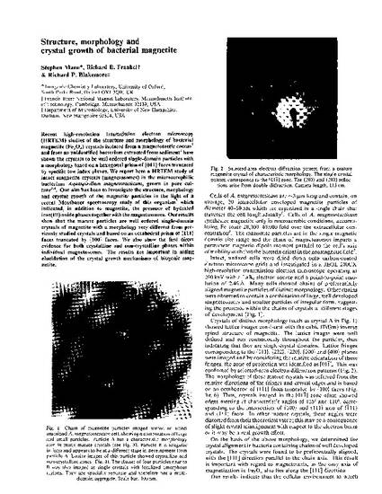
Recent high-resolution transmission electron microscopy (HRTEM) studies of the structure and morphology of bacterial magnetite (Fe3O4) crystals isolated from a magnetotactic coccus1 and from an unidentified bacterium extracted from sediment2 have shown the crystals to be well ordered single-domain particles with a morphology based on a hexagonal prism of {011} faces truncated by specific low index planes. We report here a HRTEM study of intact magnetite crystals (magnetosomes) in the microaerophilic bacterium Aquaspirillum magnetotacticum, grown in pure culture3,4. Our aim has been to investigate the structure, morphology and crystal growth of the magnetite particles in the light of a recent Mossbauer spectroscopy study of this organism5 which indicated, in addition to magnetite, the presence of hydrated iron(III) oxide phases together with the magnetosomes. Our results show that the mature particles are well ordered single-domain crystals of magnetite with a morphology very different from previously studied crystals and based on an octahedral prism of {111} faces truncated by {100} faces. We also show the first direct evidence for both crystalline and non-crystalline phases within individual magnetosomes. The results are important in aiding elucidation of the crystal growth mechanisms of biogenic magnetite.
Available at: http://works.bepress.com/rfrankel/28/
