Article
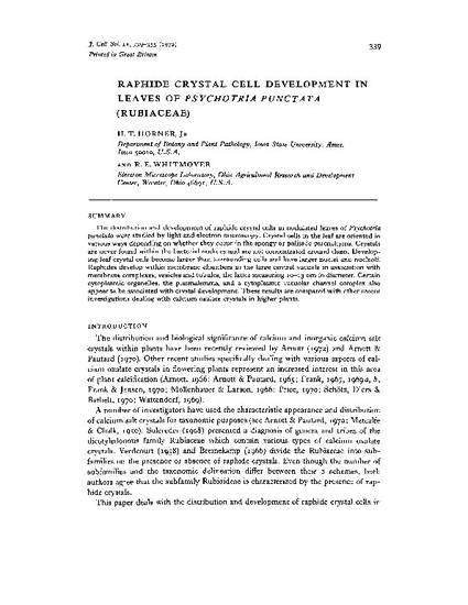
Raphide Crystal Cell Development in Leaves of Psychotria Punctata (Rubiaceae)
Journal of Cell Science
Document Type
Article
Disciplines
Publication Version
Published Version
Publication Date
1-1-1972
Abstract
The distribution and development of raphide crystal cells in nodulated leaves of Psychotria punctatawere studied by light and electron microscopy. Crystal cells in the leaf are oriented in various ways depending on whether they occur in the spongy or palisade parenchyma. Crystals are never found within the bacterial nodules and are not concentrated around them. Developing leaf crystal cells become larger than surrounding cells and have larger nuclei and nucleoli. Raphides develop within membrane chambers in the large central vacuole in association with membrane complexes, vesicles and tubules, the latter measuring 10-13 nm in diameter. Certain cytoplasmic organelles, the plasmalemma, and a cytoplasmic vacuolar channel complex also appear to be associated with crystal development. These results are compared with other recent investigations dealing with calcium oxalate crystals in higher plants.
Copyright Owner
The Company of Biologists Ltd.
Copyright Date
1972
Language
en
File Format
application/pdf
Citation Information
Harry T. Horner and R. E. Whitmoyer. "Raphide Crystal Cell Development in Leaves of Psychotria Punctata (Rubiaceae)" Journal of Cell Science Vol. 11 (1972) p. 339 - 355 Available at: http://works.bepress.com/harry-horner/45/
This article is from Journal of Cell Science 11 (1972): 339. Posted with permission.