
Tattletales and T-Bow Update 20141018
http://works.bepress.com/gmcnamara/63
Tattletales is my concept to multiplex fluorescent protein biosensors ("Tattletales", which is also the overall concept name) and multicolor FP reporters ("T-Bow" for Rainbow T-cells and tumor cells) to study the physiology of live cells.
These are based on:
1. LacI-GFP :: LacO operator synthetic tandem repeat array (Robinett et al 1996 JCB, Fig 4A, http://jcb.rupress.org/content/135/6/1685.full.pdf )
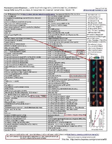
2. the plethora of fluorescent protein biosensors - see page 1 of this "63" download site.
3. the many colors, and combinations of colors (CY11.5, CyPet-YPet 90% FRET, simple color combination fusions, Steven Vogel's "V6" and "V8" fusions).
4. A need to do better than the usual overexpress FP(s) to make them easy to see (which I call "smog" in the TALE-FP and Cas9-FP papers published in 2013 and 2014).
Please see also
http://works.bepress.com/gmcnamara/42
http://works.bepress.com/gmcnamara/26
http://home.earthlink.net/~pubspectra/McNamara_20121023Tue_Tattletales_GFP_Public_Domain.jpg
The reason for the current update is this new paper:
Tanenbaum ME, Gilbert LA, Qi LS, Weissman JS, Vale RD. A Protein-Tagging System for Signal Amplification in Gene Expression and Fluorescence Imaging. Cell. 2014 Oct 8. pii: S0092-8674(14)01227-6. doi: 10.1016/j.cell.2014.09.039.
[Epub ahead of print] PubMed PMID: 25307933.
http://www.ncbi.nlm.nih.gov/pubmed/25307933
which had several nice features, though their non-optimal referencing of prior publications, such as:
* Robinett 1996 (see above)
* Lindhout 2007, 2010 - ZF-FPs
* Gross et al 2013, ora et al 2013 - FingR self regulating expression
* Yuan ... O'Farrell, TALE-Lights (especially egregious since they work at UCSF)
* other TALE-FP papers (even though smoggy)
Full references of page 2 of this "63" download pdf.
With respect to brightest colors for the T-Bow rainbow, a recent paper leads to this suggestion:
Current "best and brightest" monomeric fluorescent proteins (based on Dean and Palmer 2014) (my apology if the table does not format well online - try copy&pasting into Word or Notepad):
1. Blue mTagBFP
2. Cyan mTFP1 (“Teal”) (note: Dean and Palmer suggest mTurquoise2)
3. Green mNeonGreen or bfloGFPa1 (though each is currently wanting in terms of optimized mammalian cell expression)
alt. green UnaG … 17 kDa protein from Japanese eel … needs bilirubin … may benefit from coexpression of HO-1
4. Yellow mVenus ... more fun: Steve Vogel's V6 or V8
alt. Yellow mPapaya1 (slightly longer wavelength, dimmer) (Alternative)
5. Orange mRuby2 … 2.5x brighter than mCherry
6. Red mKate2 … or mKate S158A (our former colleague Brian Rabinovich, preferred the latter for mammalian cell imaging)
7. Far red mCardinal
8. Special C->Y CY11.5 (fusion of a cyan to a yellow FP with very high FRET)
More “colors” (excitation and emission pairs) by doing more combinations like CY11.5. Also conceivable to go “multi-FRET” such as CY11.5->mRuby2. Note that performance in mammalian cells (or other phyla!) may differe from in vitro or in Ecoli or yeast or flies rankings. Also, when authors claim "optimized expression" there is no way to know that until an even better vsion is published and proven. There are also issues of optimizing for one feature may decrease performance in others -- see mCardinal paper for example (also mEmerald, which is very bright but 50% or more molecules go "dark state" immediately - unclear from literature if thesae ever recover).
Dean KM, Palmer AE. Advances in fluorescence labeling strategies for dynamic cellular imaging. Nat Chem Biol. 2014.10: 512-23. doi: 10.1038/nchembio.1556. PubMed PMID: 24937069.
http://www.ncbi.nlm.nih.gov/pubmed/24937069
A simple way to get 2x brighter signal is to dimerize, as in Krylova et al 2013, who also localized to nucleus.
Krylova I, Kumar RR, Kofoed EM, Schaufele F. A versatile, bar-coded nuclear marker/reporter for live cell fluorescent and multiplexed high content imaging. PLoS One. 2013. 8: e63286. doi: 10.1371/journal.pone.0063286. PubMed PMID: 23691010; PubMed Central PMCID: PMC3653935.
http://www.ncbi.nlm.nih.gov/pubmed/23691010
Note: Krylova had the nice idea of dimerizing FP's with a nuclear localization signal (NLS) as a linker. However, I note that:
* localizing to Nucleolus would concentrate the signal even more ("NoLS").
* brighter FP's are now available (mCherry vs 2.5x brighter mRuby2).
* mPlum-nls-mPlum is dim ... would have been better off with mPlum-nls-mPlum-nls-mPlum (3x) or (now in 2014) mPlum-nls-mRuby2-nls-mPlum (better abosprtion using mRuby2).
* non-overlapping emission filters, in particular, adjacent bandpasses with the cutpoint between the first two and last two emission spectra, to avoid "bad photons" that appear in both emission channels. Note the longer emission channel could be "very wide bandpass" (effectively longpass).
* (possibly) somewhat shorter excitation wavelength, and wider "first emission" channel.
- Multimerization,
- Single Molecule Imaging,
- Tattletales,
- T-Bow,
- Rainbow T-cells and Tumor Cells,
- Promoter bashing,
- Fluorescent Protein Biosensors,
- Nano-Lanterns
Available at: http://works.bepress.com/gmcnamara/63/