
Presentation
Clinical Application of 3D Ultrasound in Routine Maternity Care
Society of Teachers of Family Medicine / Annual Spring Conference
(2010)
Abstract
Routine prenatal ultrasound imaging is typically performed in 2D and M-mode in Family Medicine. Pulse wave Doppler, 3D/4D, and color Doppler are modalities not commonly utilized due to the misconception that they provide minimal added information. Justification exists for the introduction of 3D, color Doppler and pulse wave Doppler in routine prenatal assessment.
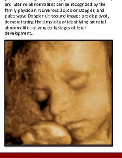
This presentation introduces first and second trimester 3D imaging techniques that allow for identification, early intervention, and surveillance of significant prenatal findings. Many fetal, placental, umbilical cord, and uterine abnormalities can be recognized by the family physician. Numerous 3D, color Doppler, and pulse wave Doppler ultrasound images are displayed, demonstrating the simplicity of identifying prenatal abnormalities at very early stages of fetal development..
Keywords
- Prenatal ultrasound,
- 3D ultrasound,
- Meningomyelocele
Disciplines
Publication Date
April 25, 2010
Location
Vancouver, CA
Citation Information
Ricardo G. Hahn, Christopher Forest, Darenie Goodman and William MacMillan Rodney. "Clinical Application of 3D Ultrasound in Routine Maternity Care" Society of Teachers of Family Medicine / Annual Spring Conference (2010) Available at: http://works.bepress.com/christopher-forest/51/