
Article
Meningomyelocele: Early Detection Using 3-Dimensional Ultrasound Imaging in the Family Medicine Center
Journal of the American Board of Family Medicine
(2010)
Abstract
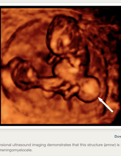
A young primigravida presented to the family medicine clinic 7 weeks pregnant. Standard 2-dimensional ultrasound at 9 weeks revealed a grossly abnormal posterior brain and 2 adjacent sonolucent structures: 2 yolk sacs versus a yolk sac and cyst. Imaging by 3-dimensional ultrasound distinguished these structures, revealing a caudal cyst with continuity of fetal tissue consistent with a meningomyelocele. To date there is no documentation in the literature of a meningomyelocele diagnosed during the first trimester of pregnancy. Identification of neural tube defects early in pregnancy offers increased options to the mother and may impact long-term fetal prognosis.
Keywords
- meningomyelocele,
- 3-dimensional ultrasound,
- neural tube defect,
- prenatal care
Disciplines
Publication Date
March, 2010
DOI
https://doi.org/10.3122/jabfm.2010.02.090226
Citation Information
Christopher Forest, Darenie Goodman and Ricardo G. Hahn. "Meningomyelocele: Early Detection Using 3-Dimensional Ultrasound Imaging in the Family Medicine Center" Journal of the American Board of Family Medicine Vol. 23 Iss. 2 (2010) p. 270 - 272 Available at: http://works.bepress.com/christopher-forest/27/