Article
Active α-macroglobulin is a reservoir for urokinase after fibrinolytic therapy in rabbits with tetracycline-induced pleural injury and in human pleural fluids
American Journal of Physiology - Lung Cellular and Molecular Physiology
(2013)
Abstract
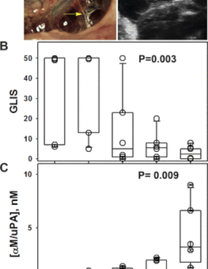
Intrapleural processing of prourokinase (scuPA) in tetracycline (TCN)-induced pleural injury in rabbits was evaluated to better understand the mechanisms governing successful scuPA-based intrapleural fibrinolytic therapy (IPFT), capable of clearing pleural adhesions in this model. Pleural fluid (PF) was withdrawn 0–80 min and 24 h after IPFT with scuPA (0–0.5 mg/kg), and activities of free urokinase (uPA), plasminogen activator inhibitor-1 (PAI-1), and uPA complexed with α-macroglobulin (αM) were assessed. Similar analyses were performed using PFs from patients with empyema, parapneumonic, and malignant pleural effusions. The peak of uPA activity (5–40 min) reciprocally correlated with the dose of intrapleural scuPA. Endogenous active PAI-1 (10–20 nM) decreased the rate of intrapleural scuPA activation. The slow step of intrapleural inactivation of free uPA (t1/2β = 40 ± 10 min) was dose independent and 6.7-fold slower than in blood. Up to 260 ± 70 nM of αM/uPA formed in vivo [second order association rate (kass) = 580 ± 60 M−1·s−1]. αM/uPA and products of its degradation contributed to durable intrapleural plasminogen activation up to 24 h after IPFT. Active PAI-1, active α2M, and α2M/uPA found in empyema, pneumonia, and malignant PFs demonstrate the capacity to support similar mechanisms in humans. Intrapleural scuPA processing differs from that in the bloodstream and includes 1) dose-dependent control of scuPA activation by endogenous active PAI-1; 2) two-step inactivation of free uPA with simultaneous formation of αM/uPA; and 3) slow intrapleural degradation of αM/uPA releasing active free uPA. This mechanism offers potential clinically relevant advantages that may enhance the bioavailability of intrapleural scuPA and may mitigate the risk of bleeding complications.
Keywords
- Fibrinolytic therapy,
- rabbit model,
- pleural injury,
- urokinase,
- α-macroglobulin,
- human
Disciplines
Publication Date
November 15, 2013
DOI
10.1152/ajplung.00102.2013
Publisher Statement
First published in the American Journal of Physiology - Lung Cellular and Molecular Physiology.
Citation Information
Azghani, A. O. (2013). Active α-macroglobulin is a reservoir for urokinase after fibrinolytic therapy. Am J Physiol Lung Cell Mol Physiol.
