Article
BioDynamic Imaging and High-Content Analysis
Journal of Biochemical Screening
(2014)
Abstract
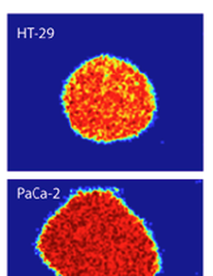
The existence of phenotypic differences in the drug responses of 3D tissue relative to 2D cell culture is a concern in high-content drug screening. Biodynamic imaging is an emerging technology that probes 3D tissue using short-coherence dynamic light scattering to measure the intracellular motions inside tissues in their natural microenvironments. The information content of biodynamic imaging is displayed through tissue dynamics spectroscopy (TDS) but has not previously been correlated against morphological image analysis of 2D cell culture. In this article, a set of mitochondria-affecting compounds (FCCP, valinomycin, nicardipine, ionomycin) and Raf kinase inhibitors (PLX4032, PLX4720, GDC, and sorafenib) are applied to multicellular tumor spheroids from two colon adenocarcinoma cell lines (HT-29 and DLD-1). These were screened by TDS and then compared against conventional image-based high-content analysis (HCA). The responses to the Raf inhibitors PLX4032 and PLX4720 are grouped separately by cell line, reflecting the Braf/Kras difference in these cell lines. There is a correlation between TDS and HCA phenotypic clustering for most cases, which demonstrates the ability of dynamic measurements to capture phenotypic responses to drugs. However, there are significant 2D versus 3D phenotypic differences exhibited by several of the drugs/cell lines.
Disciplines
Publication Date
Spring 2014
Citation Information
Ran An and David D Nolte. "BioDynamic Imaging and High-Content Analysis" Journal of Biochemical Screening (2014) Available at: http://works.bepress.com/ddnolte/4/