
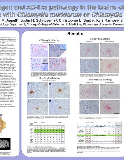
Previous research indicates BALB/c mice inoculated with Chlamydia pneumoniae (Cpn) demonstrated AD-like pathology which suggests that this mouse model is valid for studying the pathogenesis implicated in Alzheimer’s disease (AD). Studies have demonstrated that Chlamydia trachomatis (Ctr) can disseminate from its primary site of infection and plays a major role in the induction of reactive arthritis. The objectives of this lab are: (1) to identify and localize Chlamydia antigens in the brains of BALB/c mice infected with C. muridarum and (2) to determine if infection with C. muridarum induces AD-like pathology comparable to Cpn. Using mouse adapted respiratory isolates of C. muridarum, we investigated whether C. muridarum disseminated from the respiratory tract to the brain. Mice were intranasally infected with plaqued C small Weiss (CSW) or plaqued mouse pneumonitis Weiss (MoPn Weiss). Brain tissue was isolated at 2 months post-infection. Serial sections from brains infected mice were analyzed for amyloid or Chlamydia antigens. Preliminary analysis of brain tissue demonstrated no detectable difference in C. muridarum antigen between mice receiving 1 x10⁵ IFU and mice receiving 1 x101 IFU, whereas a small but detectable difference was identified in amyloid-specific labeling between these two experimental groups. In contrast, prominent Chlamydia-specific labeling was identified in the brains of Cpn-infected mice as well as substantial amyloid deposition at 2 months p.i.. These data suggest that, relative to Cpn AR-39 infection, C. muridarum infection is a weaker stimulus for inflammation, resulting in decreased amyloid deposition in the brains of BALB/c mice.
Available at: http://works.bepress.com/c_little/11/